Inferior view of the skull mandible removed – Unveiling the intricate details of the skull’s inferior view, this comprehensive exploration delves into the anatomical structures, muscle attachments, foramina, and canals that define this region. From its clinical significance to its diagnostic applications, this investigation illuminates the essential role of the inferior view in various medical fields.
Inferior View of the Skull (Mandible Removed): Inferior View Of The Skull Mandible Removed

The inferior view of the skull, with the mandible removed, provides a comprehensive perspective of the skull’s base. This view reveals essential anatomical landmarks and structures that play a crucial role in various medical fields.
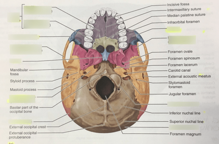
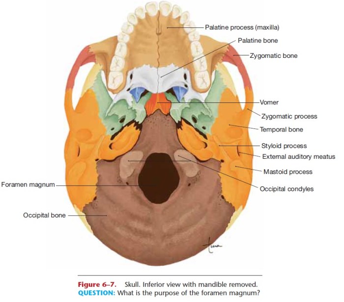
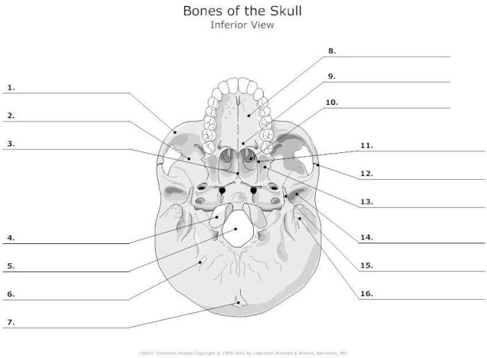
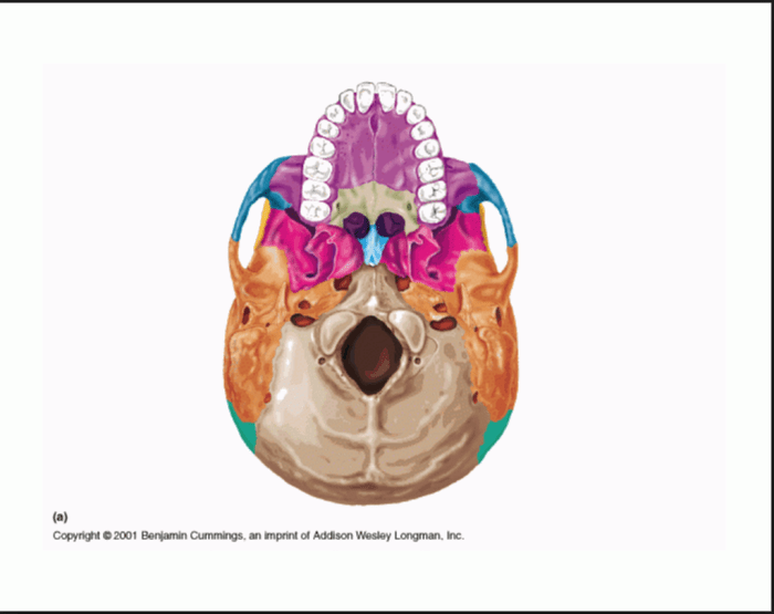
The inferior surface of the skull is composed of several bones, including the occipital bone, temporal bones, and sphenoid bone. These bones articulate to form the foramen magnum, which serves as the passageway for the spinal cord. Additionally, the inferior view exhibits numerous foramina and canals that transmit nerves and blood vessels.
Anatomical Structures, Inferior view of the skull mandible removed
| Bone | Description |
|---|---|
| Occipital Bone | Forms the posterior portion of the skull; contains the foramen magnum |
| Temporal Bones | Located on either side of the occipital bone; house the auditory organs and balance apparatus |
| Sphenoid Bone | Located at the base of the skull; contains the sella turcica, which houses the pituitary gland |
| Foramen | Description |
|---|---|
| Foramen Magnum | Large opening at the base of the skull; transmits the spinal cord |
| Jugular Foramen | Paired foramina on either side of the foramen magnum; transmits the jugular vein, glossopharyngeal nerve, vagus nerve, and accessory nerve |
| Hypoglossal Canal | Foramen located anteriorly to the jugular foramen; transmits the hypoglossal nerve |
Muscle Attachments
- Rectus Capitis Posterior Minor: Originates from the posterior arch of the atlas; inserts onto the occipital bone; extends the head
- Rectus Capitis Posterior Major: Originates from the spinous process of the axis; inserts onto the occipital bone; extends the head
- Obliquus Capitis Inferior: Originates from the spinous process of the axis; inserts onto the transverse process of the atlas; rotates the head
- Obliquus Capitis Superior: Originates from the transverse process of the atlas; inserts onto the occipital bone; rotates the head
Foramina and Canals
| Foramen | Function | Structures Transmitted |
|---|---|---|
| Foramen Ovale | Transmits the mandibular nerve, accessory meningeal artery, and lesser petrosal nerve | |
| Foramen Spinosum | Transmits the middle meningeal artery and meningeal branch of the mandibular nerve | |
| Jugular Foramen | Transmits the jugular vein, glossopharyngeal nerve, vagus nerve, and accessory nerve |


